 Diastole:
In order to grasp the concept of measuring and interpreting hemodynamic values, it is important to understand how blood flowing through the heart is related to the cardiac cycle.
Fluid moves from high pressure to lower pressure.
Blood within the cardiovascular system adheres to this rule as evidenced by the direction of blood flow. The higher pressure generated by the left heart produces a gradient which moves blood from the left heart, through the body tissues to collect in the right side of the heart.
Diastole:
Systole:
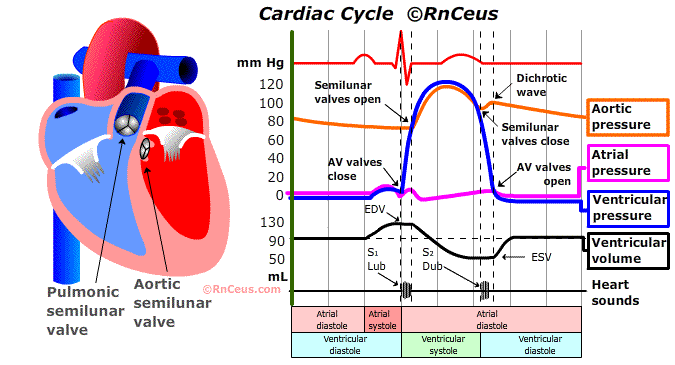
When the left ventricle (LV) contracts, it generates a systolic blood pressure of 100-140 millimeters of Hg (mm Hg).
The pressure of blood within the right atrium is the central venous pressure (CVP).
The blood pressure of the vena cavae is similar to the CVP because there are no valves or flow obstructions between the vena cavae (VC) and the RA. The VC and heart's right side can be viewed as one chamber with a contractile portion at the distal end. The CVP averages between 2-6 millimeters of mercury (mm Hg).
During right ventricular (RV) diastole, the pressure within the RV is between 0-5 mm Hg.
Elasticity and compliance of the ventricular myocardium help generate a lower intraventricular pressure. Lower intraventricular pressure, aided by atrial systole, causes blood to flow across the open atrioventricular AV valve.
Right ventricular systolic pressure is usually from 20-30 mm Hg.
This exceeds the right atrial pressure. The pressure gradient applies greater pressure to the ventricular side of the AV valve, which causes it to close.
The pulmonary artery (PA) pressure, prior to systole, is normally 8-12 mm Hg.
During RV systole the PA pressure will rise to equal the RV pressure, usually 20-30 mm Hg. The momentary systolic PA pressure of 20-30 mm Hg is quickly dissepated by the compliance of the pulmonary vascular bed and the PA returns to a diastolic pressure of 8-12 mm Hg.
Blood leaves the pulmonary vasculature at about 4-12 mm Hg, moving into the pulmonary veins.
The pulmonary veins empty directly into the left atrium . Elasticity and compliance of the ventricular myocardium help generate a slightly lower intraventricular filling pressure. Lower intraventricular pressure, aided by atrial systole, causes blood to fill the left ventricle and stretch the ventricular myofibril to the left ventricle end-diastolic volume (LVEDV).
The LVEDV provides the cardiac myofibril stretching force (preload) that operates the Frank-Staring mechanism. As Guyton put it, "When the cardiac muscle becomes stretched an extra amount, as it does when extra amounts of blood enter the heart chambers, the stretched muscle contracts with a greatly increased force, thereby automatically pumping the extra blood into the arteries." Conversely, a reduction of intravascular volume that reduces venous return and LVEDV will reduce stroke volume and cardiac output.
LV systole generates 100-140 mm Hg.
Aortic pressure during diastole is usually 60-90 mm Hg (afterload). This is the pressure the left ventricle must overcome to open the aortic valve and eject blood into the aorta. The pressure gradient of systolic 100-140/60-90 mm Hg drives blood into the aorta and onward to the rest of the body. The cycle is complete.
References
Guyton A.C. (1986). Textbook of Medical Physiology. London: W. B. Saunders
Tkacs, Nancy C., et al. (2020). Advanced Physiology and Pathophysiology: Essentials for Clinical Practice. Springer Publishing Company.
© RnCeus.com